

4DMedical CE Mark: The Lung Imaging Platform That Makes Routine CT Scans See More
Every year, tens of millions of chest CT scans are performed across hospitals worldwide. They are among the most common and clinically useful diagnostic tools in medicine, capable of revealing tumours, infections, structural abnormalities, and the damage wrought by decades of lung disease. What they have not been able to do, at least not routinely, is tell a clinician how the lung is actually functioning. A structural image shows what the lung looks like.
It does not show how air moves through it, which regions are ventilating properly, where blood flow is compromised, or how disease is progressing at a regional, lobar level. The gap between what a CT can see and what a clinician needs to know has been one of the defining limitations of respiratory medicine for decades. Melbourne-based 4DMedical was built to close it.
On March 30, 2026, 4DMedical announced that its flagship technology, CT:VQ, had received CE Mark certification for commercial use across the European Union. On the same day, the company confirmed it had secured AU$83 million in a private placement from institutional investors, capital specifically directed at accelerating European commercial deployment.
With FDA 510(k) clearance already in place and active deployments now running at six of the most prestigious academic medical centres in the United States, including Stanford, Cleveland Clinic, Mayo Clinic, and UC San Diego Health, 4DMedical is now commercially cleared across both the world’s largest and most sophisticated healthcare markets simultaneously.
The Problem With How We Have Always Imaged Lungs
Pulmonary disease is fundamentally functional. The clinical reality of conditions including COPD, pulmonary embolism, idiopathic pulmonary fibrosis, chronic thromboembolic pulmonary hypertension, and asthma is defined by abnormalities in how air moves through the lung and how blood perfuses it. A structural image can reveal the consequences of these abnormalities, thickened walls, emphysematous destruction, fibrotic scarring, but it cannot reveal the functional reality in real time, cannot quantify regional ventilation, and cannot map perfusion without injecting contrast agents or exposing patients to radioactive tracers.
The conventional alternative to CT for functional lung imaging is nuclear medicine ventilation-perfusion scanning, or VQ scanning, which requires radioactive tracers, specialised nuclear medicine infrastructure, dedicated scheduling, trained personnel, and often a separate patient visit. Access to nuclear VQ scanning is constrained globally: not every hospital has nuclear medicine capacity, radiotracer supply can be unreliable, and the operational complexity of the procedure limits its use precisely where respiratory disease burden is highest.
Approximately 400,000 nuclear VQ scans are performed annually across the EU alone, according to 4DMedical’s estimates, representing a meaningful clinical need that existing infrastructure is struggling to meet.
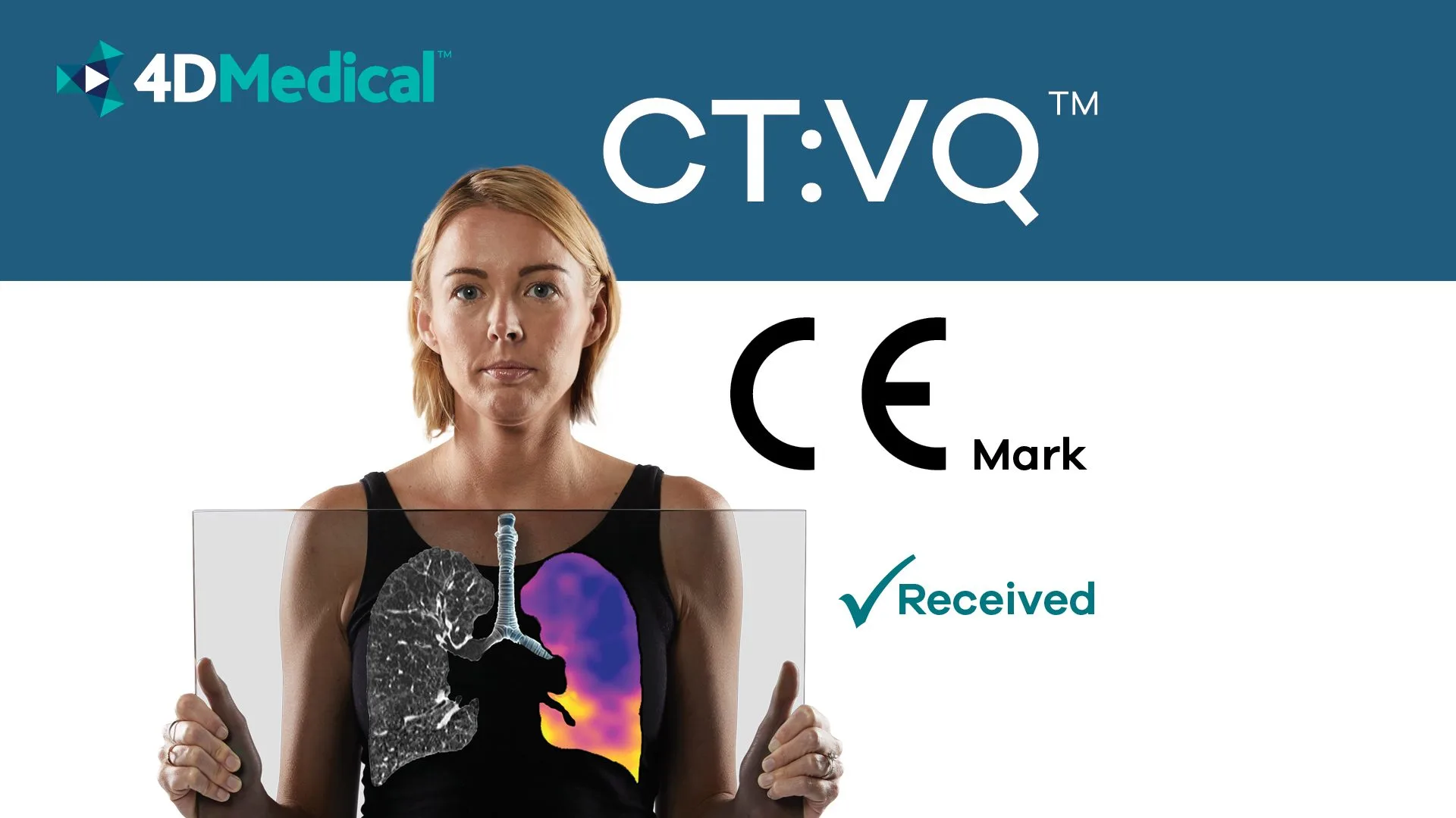
CT:VQ: Maximum Contrast, No Injections
CT:VQ is 4DMedical’s answer to this structural gap in respiratory diagnostics. It is the world’s first and only non-contrast ventilation-perfusion imaging technology, delivering quantitative functional lung insights from a routine, standard chest CT scan without contrast agents, radioactive tracers, or any additional specialised equipment.
The software processes existing CT data and generates colour-coded ventilation and perfusion maps registered directly to the CT anatomy, giving clinicians a lobar view of how each region of the lung is ventilating and perfusing, alongside quantitative metrics including total lung volumes, lobar distribution comparisons, and right-to-left lung analysis.
- 14,500 CT scanners installed across the US that CT:VQ can run on with no new hardware
- 4,00,000 Nuclear VQ scans performed annually in the EU that CT:VQ can supplement or replace
- 450M+ People in the EU now covered by 4DMedical’s CE Mark-enabled commercial deployment
The clinical applications are broad. Pulmonologists use CT:VQ to support pulmonary embolism workups, assess chronic thromboembolic pulmonary hypertension, detect regional ventilation-perfusion abnormalities, plan bronchoscopic lung volume reduction procedures, and conduct pre-operative assessments and longitudinal disease monitoring. Radiologists receive structured quantitative outputs delivered directly to PACS via DICOM Structured Reporting, adding functional interpretation to non-contrast chest CTs without disrupting existing workflow.
For hospital administrators, the proposition is equally compelling: expanding access to functional lung imaging across an existing CT fleet with no new hardware investment and no requirement for nuclear medicine infrastructure, while also generating reimbursement through Category III CPT codes in the US market.
The clinical evaluation of CT:VQ used three complementary approaches to validate performance against the existing gold standard of SPECT VQ scanning. Reader performance studies showed good to excellent agreement with SPECT VQ across all lung zones, with Kendall’s tau by zone ranging from 0.702 to 0.765, exceeding the pre-specified 0.40 target for the FDA submission. Additional analysis showed CT:VQ perfusion heterogeneity metrics demonstrating a stronger association with DLCO than comparable SPECT measures in the submission dataset.
“CE Mark certification for CT:VQ opens access to one of the world’s largest and most sophisticated healthcare markets. The clinical need is universal. The limitations of nuclear VQ scanning exist across healthcare systems globally.” – Andreas Fouras, Managing Director, CEO and Founder, 4DMedical

A Platform Wider Than a Single Product
CT:VQ is 4DMedical’s commercial flagship, but it sits within a considerably broader software platform built on the company’s patented XV Technology, the proprietary algorithmic foundation enabling four-dimensional, quantitative respiratory imaging. The full product suite spans the spectrum of cardiopulmonary assessment, with each tool designed to extract additional clinical value from existing CT and X-ray infrastructure rather than requiring new modalities.
- IQ-UIP: AI-powered deep learning tool achieving 90.2% sensitivity and 91.5% specificity in detecting UIP patterns associated with idiopathic pulmonary fibrosis. FDA-cleared in 2024 as a Breakthrough Device. Cuts average diagnostic delay from two years in IPF patients.
- XV LVAS: 4DMedical’s original lung ventilation analysis system based on XV Technology. Quantitatively reveals patterns of regional lung function in a four-dimensional and highly visual manner from X-ray imaging.
- CT LVAS: Lung ventilation analysis from CT imaging. Generates regional ventilation outputs to support assessment of obstructive and restrictive lung disease alongside structural CT findings.
- PHA: Pulmonary hypertension assessment tool providing quantitative cardiac and vascular measurements from routine CT to support PH evaluation without dedicated cardiac imaging.
- LDAi and LDAf: Inspiration and functional lung density analysis tools delivering quantitative measures relevant to emphysema assessment and tracking disease progression longitudinally.
- CAC: Coronary artery calcification quantification from thoracic CT, providing a cardiovascular risk marker from scans already being performed for respiratory indications.
The logic threading all these products together is consistent: routine chest CT contains far more clinically actionable information than conventional reporting extracts. 4DMedical’s software platform is the mechanism for extracting it, quantifying it, and delivering it in forms that integrate into clinical workflows without adding appointments, procedures, or specialist dependencies.
The Lung Health Imaging Panel, which bundles LDAi, CAC, IQ-UIP, and lung cancer screening tools into a single comprehensive output from a thoracic CT, exemplifies this philosophy at its most ambitious: one scan, many insights, no additional patient burden.
4DMedical European Expansion and Beyond
The CE Mark of CT:VQ™ opens a market of more than 450 million people with a highly developed hospital-based imaging infrastructure and a substantial installed base of CT scanners. Europe’s research institutions and academic centres have also been among the most active contributors to the clinical literature on advanced lung imaging, making the region not only a commercial opportunity but a natural environment for the kind of evidence generation and institutional validation that drives adoption in radiology and pulmonology.
4DMedical has described its European expansion strategy as built around working with leading hospitals and clinicians on adoption, evaluation, and research initiatives, following the model established by its US deployments at Stanford, Cleveland Clinic, Mayo Clinic, the University of Miami, UC San Diego Health, and the University of Chicago Medicine. The AU$83 million raised alongside the CE Mark announcement provides the capital to execute that strategy at pace.
For a company whose mission is to improve outcomes for the hundreds of millions of people worldwide living with lung disease, the combination of regulatory clearance across two major markets, a validated and growing clinical evidence base, and institutional capital to fund the commercial buildout represents a convergence of the conditions that medical technology companies work toward for years before seeing arrive simultaneously.